Ridge-Preservation Open membrane technique with Geistlich Bio-Gide® and Geistlich Bio-Oss®, implant Placement. A case report:
CASE
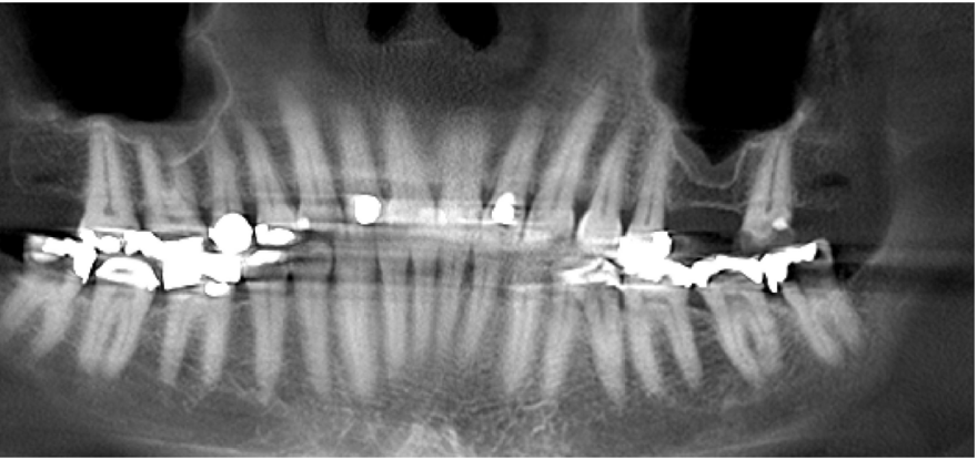
37years old female
First visit 2016.10
No medical history
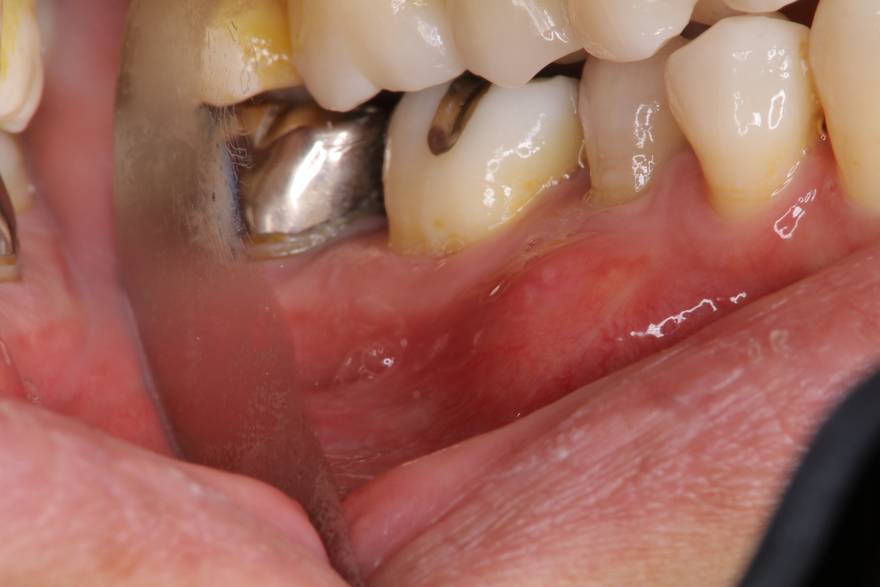
2016.12.
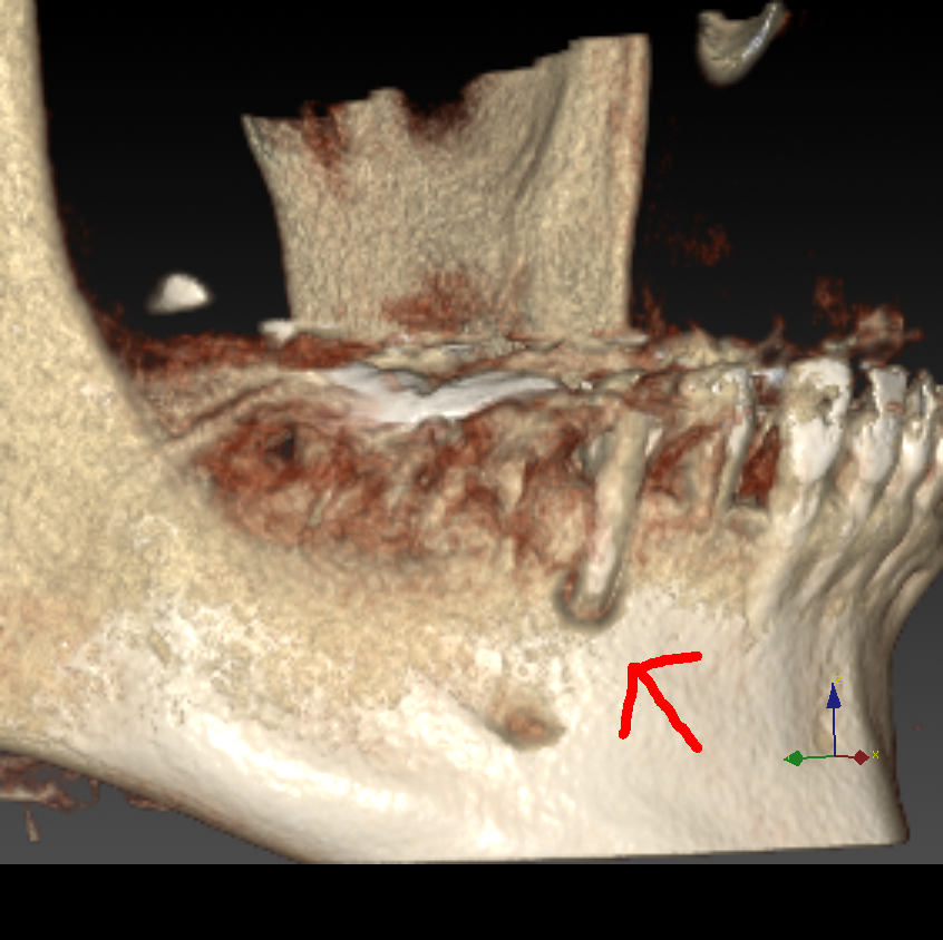
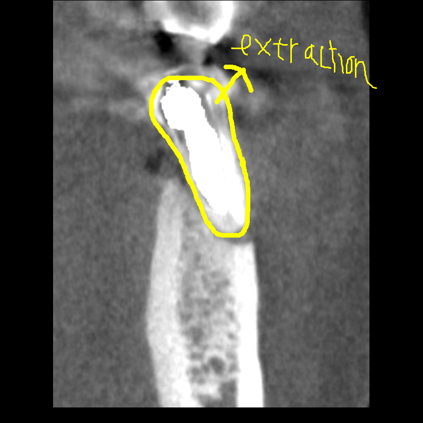
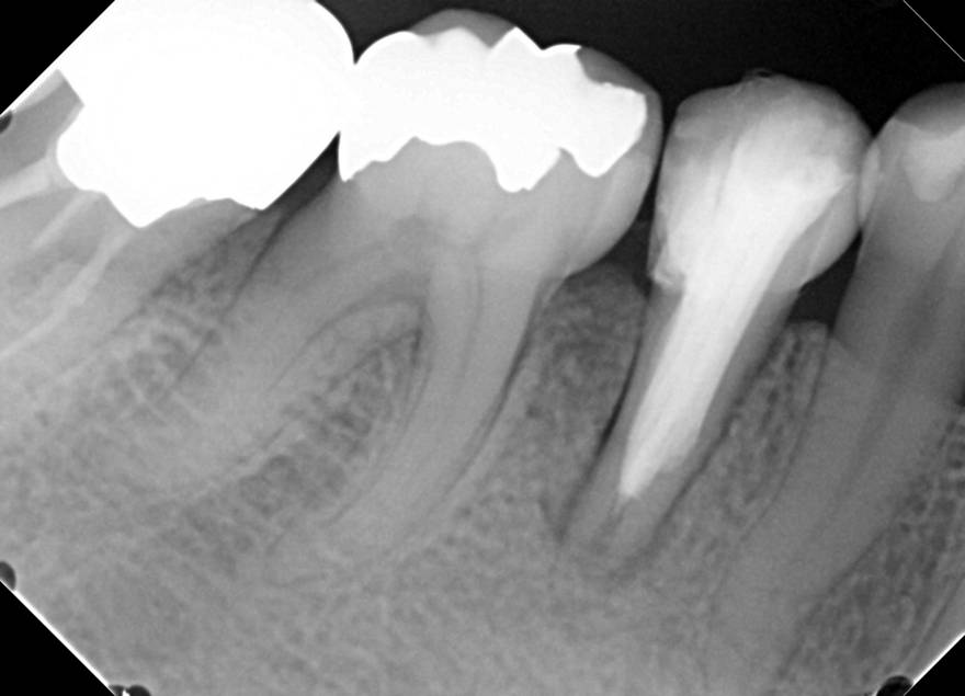
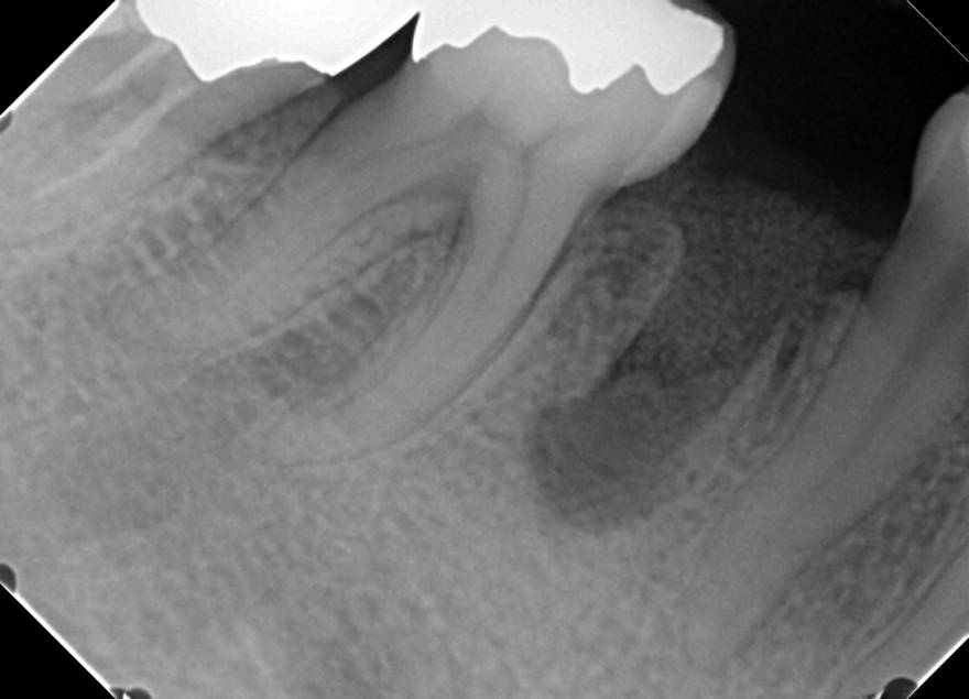
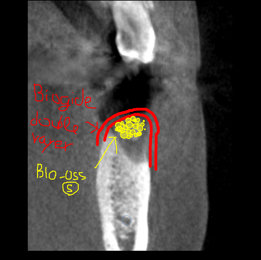
45 extraction, and debridement of the inflammatory tissue.
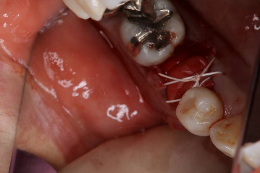
Filling with Geistlich Bio-Oss® (small granules 0.25-1mm)
Buccal bone wall replaced by Geistlich Bio-Gide® (13×25mm) double-layer technique
Geistlich Bio-Gide® is sutured with a modified cross-mattress suture with 4-0 PTFE, allowing optimal adaptation between the borders of the soft tissues and the collagen matrix
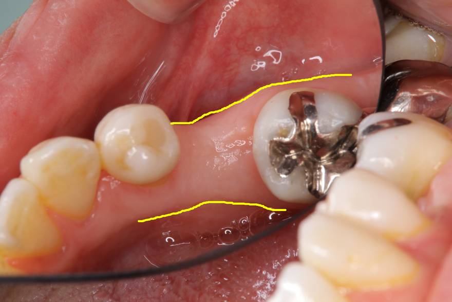
Situation after 17days after surgery
Note the epithelium has become stretched
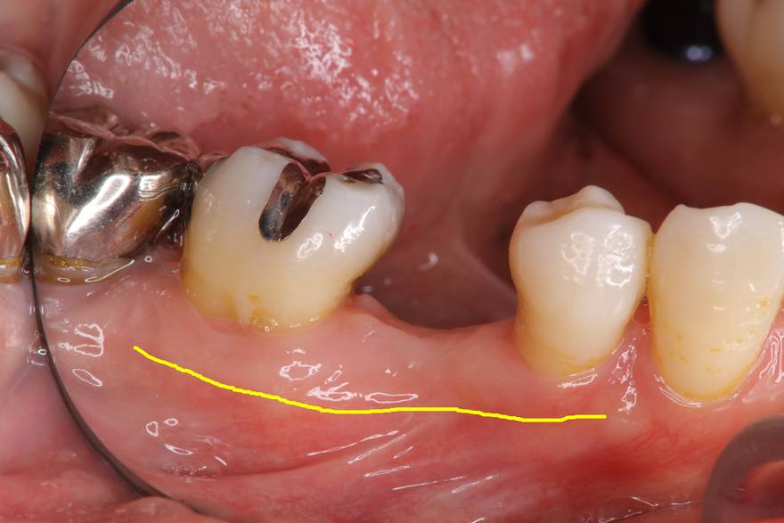
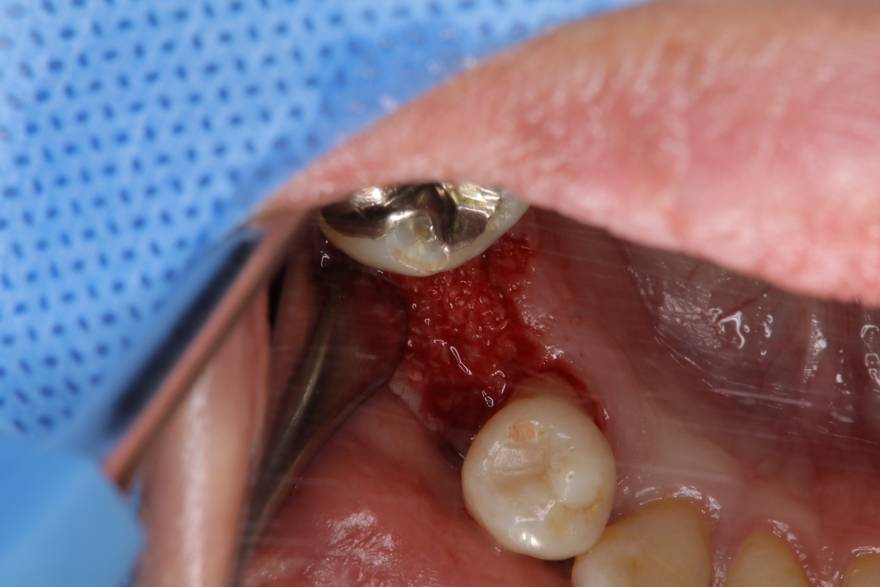
2017.7
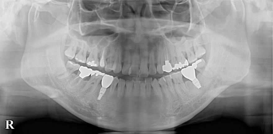
Clinical situation after 8 months of healing,
Maintains the alveolar volume
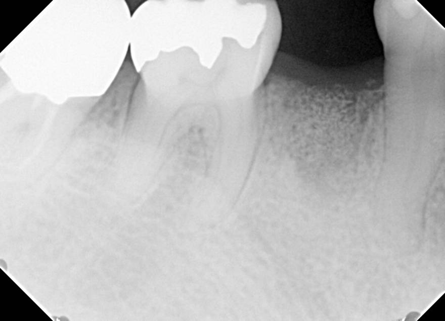
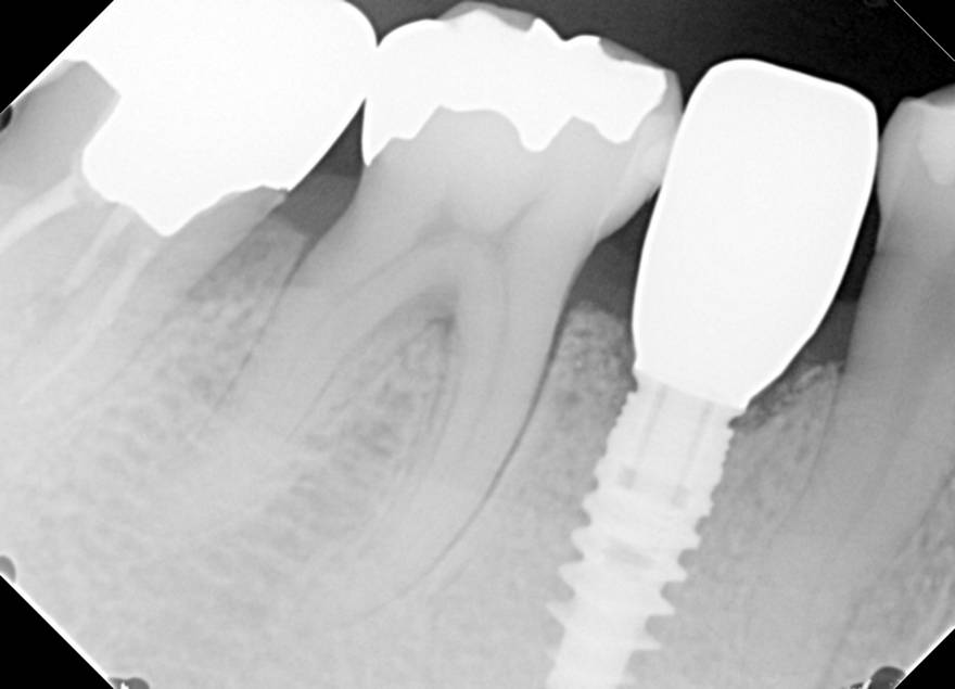
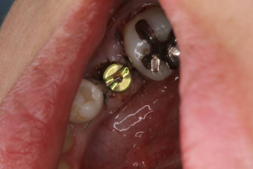
45 implant placement(Nobel active Rp4.3*11.5mm)
primary stability is very good
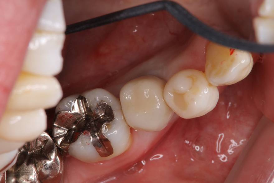
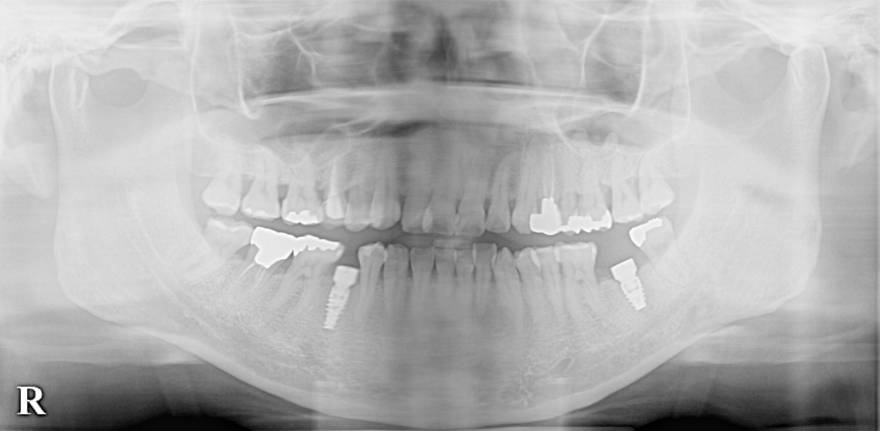
Final prosthetic and well maintained the alveolar volume
3-D bone augmentation using a CAD/CAM customized titanium mesh in conjunction with autogenous bone and bovine bone mineral granules
A 75-year old and systemically healthy female came to our attention requiring a fixed rehabilitation in the bilateral mandibular edentulous areas, from teeth 35 to 38 and from 45 to 48. The clinical and radiological evaluation showed vertical and horizontal bone atrophies. Based on a computed tomography of the defect (CBCT) carried out before the surgical procedure with the aid of a radiopaque diagnostic template, DICOM scans of the three-dimensional models are reproduced in a very precise way. This makes it possible to produce a precise and accurate three-dimensional titanium scaffold which on its outer part represents the final shape/profile of the new and ideal alveolar ridge, while in the inner part containing and stabilizing the grafting material. This latter is generally represented by a mixture of autogenous bone chips and bovine bone mineral. The mesh was covered with a Geistlich Bio-Gide® in order to optimize the barrier effect.
Multiple buccal recession defects ranging 2-5 mm were noted by teeth #11-14 with a minimal amount of keratinized tissue on the buccal of #14. Bone levels were within normal limits with no loss of interproximal tissue observed. These recession defects are classified as Miller Class I recession defects. Typically, 100% root coverage is expected for recession defects of this type. The treatment was complete root coverage of the recession defects and augmentation of the width of attached keratinized tissue by tooth #14. Complete root coverage and an increase in the zone of keratinized tissue was obtained and a dento-gingival complex that is amenable to long-term health and stability was achieved. The patient was spared from the inevitable morbidities associated with a sub-epithelial connective tissue graft from a palatal donor site.
A patient with a progressive gum recession which had led to compromised esthetics and sensitivity. The teeth had 3-4 mm of gingival recession on the buccal surface with a sufficient zone of
keratinized gingiva. The patients' main priorities were to improve
esthetics and reduce/eliminate root sensitivity. Treatment goals for this case were to obtain complete root coverage, increase soft-tissue thickness, and reduce/eliminate cervical sensitivity. Using Geistlich Fibro-Gide® a 100% root coverage has been obtained
and the patients' chief complaints of esthetics and sensitivity have been addressed.